Stereology is the science of obtaining quantitative information about three dimensional structures from lower dimensional observations. In practical terms, it allows researchers to estimate properties such as volume, surface area, length, number and spatial distribution by studying sections, slices or images of a larger object. It is widely associated with microscopy because many biological and material samples cannot be observed completely in three dimensions without being cut, scanned or otherwise sampled. A researcher examining a thin tissue section sees only a small part of the original structure. Stereology provides a mathematical framework for turning those partial observations into defensible estimates of the whole.
The field matters because visual inspection can be misleading. A large structure may appear frequently in sections simply because it occupies more space. Large cells may have a greater chance of being intersected than small cells. Long fibres may appear as many disconnected profiles. Counting what is visible on a microscope slide therefore does not automatically reveal how many objects exist in the original tissue. Stereological methods address these problems by combining geometrical probability with controlled sampling. The aim is not to reconstruct every object individually, but to estimate population level properties without systematically favouring certain shapes, sizes or positions.

Stereology Is About Inference Rather Than Reconstruction
Stereology is sometimes confused with three dimensional reconstruction, but the two methods ask different questions. Reconstruction attempts to recreate the geometry of an individual structure from a series of images or sections. A researcher might trace a neuron through consecutive tissue slices and use software to build a three dimensional representation. Stereology does not necessarily attempt to reconstruct that neuron. Instead, it may estimate the total number of neurons in a brain region, the total length of blood vessels, or the proportion of tissue occupied by a particular cell type.
This distinction makes stereology particularly useful when a complete reconstruction would be impractical. A biological organ can contain millions or billions of relevant structures. Recording every cell, capillary or membrane directly would require enormous amounts of time and data. Stereological sampling allows a much smaller number of observations to represent the larger population, provided that the sampling procedure is properly designed.
The logic resembles statistical sampling in other sciences. Measuring every member of a population is unnecessary if a representative sample can produce an accurate estimate. Stereology adds geometrical rules because the objects are embedded in space. The way a plane cuts through a sphere, fibre or irregular particle affects what appears in the sample. Ignoring that geometry introduces systematic bias.
The Central Problem Is Moving From 2D to 3D
Most stereological problems begin with a dimensional mismatch. The object being studied exists in three dimensions, but the observations may be two dimensional. A tissue section has area but very little thickness. A microscope image may show points, lines and profiles even though the original structures are volumes, surfaces and fibres.
Imagine cutting an orange into thin slices. A slice through the centre produces a large circular profile, while a slice near the edge produces a smaller one. If only one slice were available, the size of that circle would not tell you the exact volume of the orange unless you already knew where the cut had been made. The problem becomes harder when the object has an irregular shape or when many objects overlap within the same sample.
Stereology solves this by designing the sampling process around probability. Rather than trying to guess the three dimensional form from one convenient section, it uses appropriately selected sections and geometrical test systems. The relationship between the observed sample and the underlying structure can then be described mathematically.
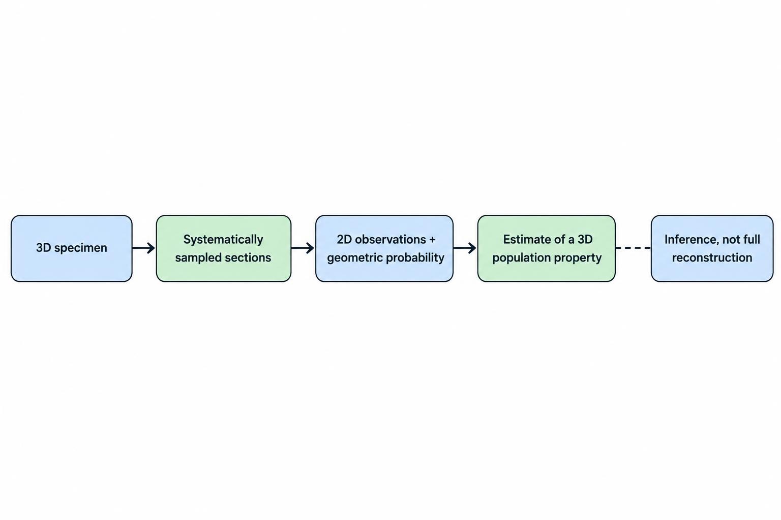
This is why sampling design is more important in stereology than the visual sophistication of the microscope image. A beautifully resolved but badly sampled image can produce a precise measurement of the wrong thing.
Early Stereology Developed From Geometrical Probability
The intellectual roots of stereology lie in geometry, probability and microscopy. Scientists had long recognised that sections through solid objects contain information about their three dimensional structure. Metallurgists studied polished surfaces to estimate the properties of grains and particles inside metals, while anatomists examined tissue sections to understand biological organisation.
The difficulty was converting observation into quantitative measurement. Researchers needed to know when a two dimensional profile could represent a three dimensional quantity and when the relationship depended too heavily on object shape or orientation.
One important line of development involved geometrical probability. If a set of test points is placed randomly over a section, the proportion of points hitting a structure can be related to the fraction of volume occupied by that structure. If test lines intersect surfaces under appropriate sampling conditions, the number of intersections can be related to surface density. These relationships made it possible to measure quantities that seemed inaccessible from sections alone.
By the twentieth century, these principles were being organised into a distinct quantitative discipline. Modern stereology developed further as researchers introduced methods designed specifically to avoid assumptions about object size, shape and orientation.
Unbiased Sampling Is the Foundation of Stereology
A stereological estimate is only as defensible as the sampling scheme used to obtain it. If some parts of a specimen are more likely to be sampled than others without a mathematical reason for that difference, the final result can be biased.
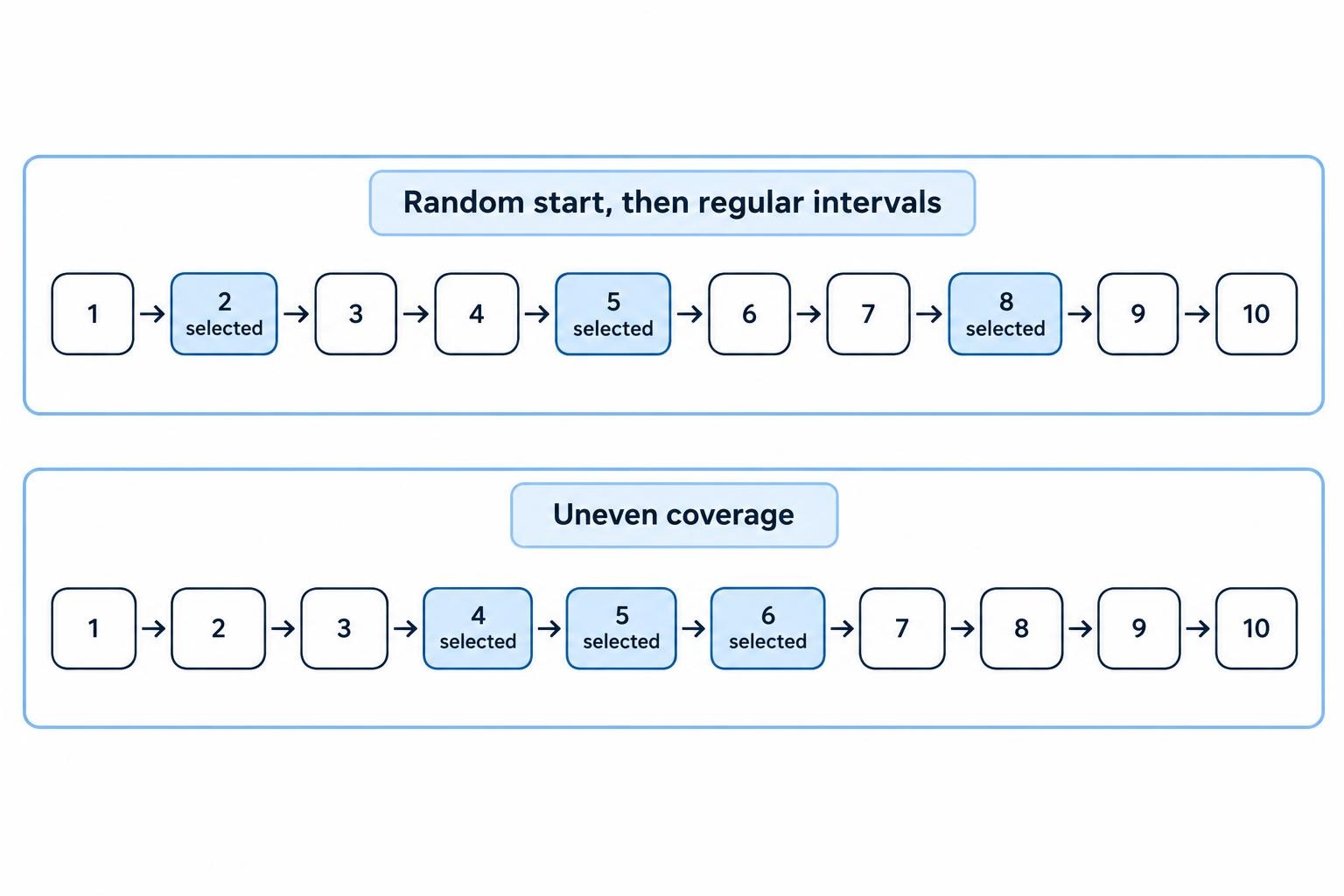
Random sampling is therefore central. In simple random sampling, every potential sampling location has an equal chance of selection. In practice, however, stereological studies commonly use systematic random sampling. A random starting location is chosen, after which sections or fields are sampled at regular intervals. This combines the protection of randomisation with the efficiency of systematic spacing.
Suppose a brain region is cut into 200 serial sections. Examining every section might be unnecessary. A researcher could randomly select one of the first ten sections and then analyse every tenth section thereafter. The starting point is random, but the rest of the sample is evenly distributed through the tissue.
This method usually provides better coverage than selecting the same number of sections independently at random because systematic sampling spreads the observations throughout the entire structure.
The Sampling Fraction Must Be Known
Stereological estimation depends on knowing how much of the original material was sampled. If one section out of every ten is examined, the section sampling fraction is one tenth. If only one quarter of each selected section is analysed, another sampling fraction is introduced.
These fractions allow observed counts to be expanded into estimates of totals. If a known fraction of the structure has been sampled using an unbiased procedure, the total population can be estimated from the number observed.
The calculation is conceptually simple but depends on careful record keeping. Researchers need to know which sections were included, how sampling frames were placed and how much tissue was actually evaluated. Changing the sampling interval halfway through an experiment or selecting visually interesting regions can destroy the relationship between sample and population.
This is why stereology is not simply a collection of counting formulas. It is a sampling discipline. The formulas work because the sampling probabilities are controlled.
Orientation Can Be as Important as Position
Some stereological measurements depend not only on where a section is taken but on how it is oriented.
Consider a bundle of parallel fibres. A section cut perpendicular to the fibres may show many roughly circular profiles. A section cut parallel to them may show long lines. Both images come from the same structure, yet the appearance is completely different.
If the quantity being estimated is sensitive to orientation, sections may need to be isotropic, meaning all spatial directions are represented without systematic preference. Researchers can achieve this through specialised randomisation procedures.
Other measurements do not require isotropic sections. Volume estimation through point counting, for example, can often be performed on ordinary systematic sections. Surface area or length measurements can be more sensitive to orientation and may require more careful preparation.
Recognising which measurements depend on orientation prevents a common stereological mistake: applying a valid counting rule to a sample whose geometry violates the assumptions behind that rule.
Point Counting Is One of the Simplest Stereological Methods
Point counting is widely used to estimate volume fractions. A grid containing test points is placed over sampled sections, and the researcher records how many points land on the structure of interest.
If 30% of the test points fall on a particular tissue component, that component is estimated to occupy approximately 30% of the reference volume, assuming appropriate sampling.
The power of the method lies in its simplicity. The researcher does not need to trace every boundary or calculate the exact area of each visible profile. The points provide a probability sample of the area, which can then represent the volume fraction.
More points generally reduce sampling uncertainty, although the relationship is not infinitely efficient. Beyond a certain point, analysing more independent fields may provide more information than placing an extremely dense grid over the same field.
Point counting is common in histology because many biological questions concern proportions. A researcher might estimate the fraction of liver occupied by fat, the proportion of tumour tissue containing necrosis, or the fraction of lung occupied by air spaces.
The Cavalieri Principle Is Used to Estimate Volume
The Cavalieri principle provides a straightforward method for estimating the total volume of an irregular object from a series of parallel sections.
The basic idea is to sample sections through the object at a known interval, measure the area of the structure on each section and combine those areas with the distance between sections. In practice, area is often estimated by point counting rather than tracing every boundary.
If sections are sampled systematically from a random starting position, the method can provide an unbiased estimate of total volume regardless of the object’s irregular shape.
This is useful in biology because organs and brain regions are rarely simple geometric solids. Calculating their volume using assumptions about spheres or cylinders would introduce unnecessary model dependence.
A stereological volume estimate instead relies on the observed sections themselves. The organ can bulge, narrow or change shape along its length without invalidating the method.
The same logic is useful in materials science when measuring irregular particles, pores or phases distributed through a specimen.
Counting Objects Is Harder Than Counting Profiles
Estimating the number of cells or particles creates one of the most important stereological problems. A two dimensional section displays profiles, not whole objects.
A large cell has a greater chance of being cut by a section than a small cell. If visible profiles are simply counted, large objects tend to be overrepresented. One object can also appear in more than one adjacent section, leading to repeated counting.
This is why older profile based counting methods could produce biased estimates when object size or shape differed between experimental groups.
Modern stereology addresses the problem by counting objects according to events that occur within a known three dimensional sampling space rather than simply counting every visible cross section.
The disector is the most important example. It estimates numerical density or total number without requiring assumptions about cell size, shape or orientation.
The Disector Changed Stereological Counting
The disector uses information from two parallel sections or from a three dimensional optical sampling volume. An object is counted when it appears according to a predefined rule in one plane but not the comparison plane, or when a designated feature first comes into focus while moving through the sampling volume.
The important point is that the probability of being counted depends on the object’s presence within the sampling volume rather than the size of its profile.
This makes the method much less sensitive to differences in cell diameter. A larger cell is not counted repeatedly simply because it intersects more sections.
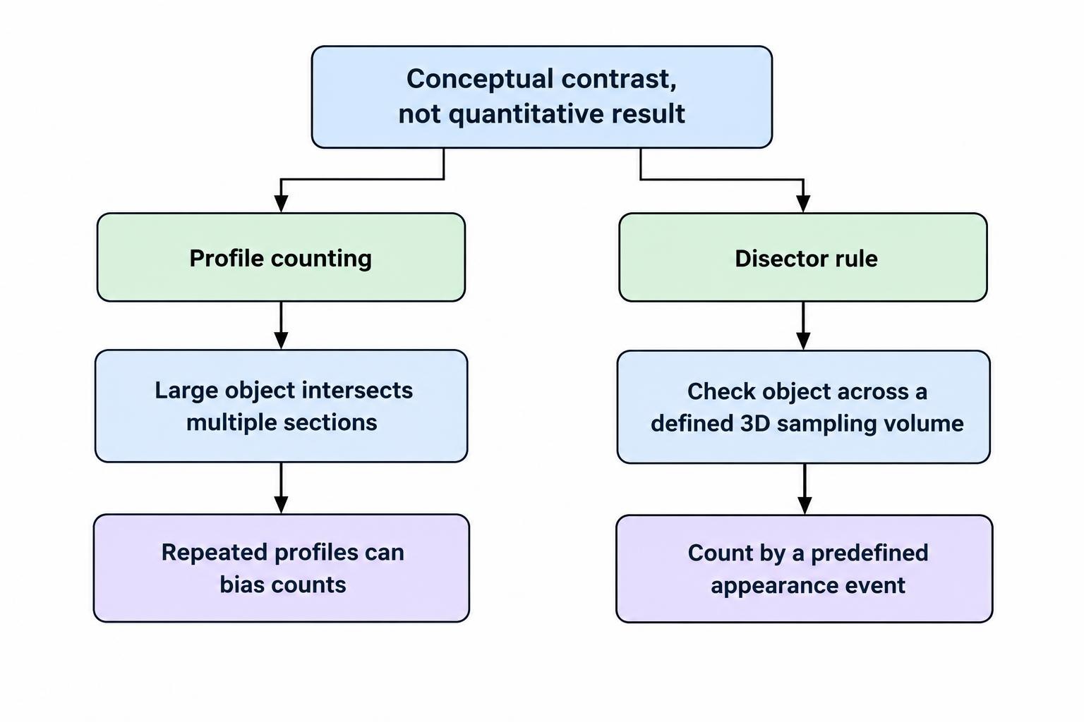
Two broad versions are commonly discussed. A physical disector uses two actual sections separated by a known distance. An optical disector moves through the depth of a relatively thick section using a microscope capable of resolving objects at different focal levels.
The optical approach has become common in neuroscience and histology because it allows researchers to sample cells in three dimensions without physically producing a separate comparison section for every counting frame.
The Optical Fractionator Estimates Total Number
The optical fractionator combines the optical disector with systematic random sampling to estimate the total number of objects in a defined structure.
The method is particularly well known in neuroscience, where researchers may want to estimate the number of neurons in a brain region.
Rather than calculating numerical density first and then multiplying by an estimated volume, the fractionator samples a known fraction of the total structure directly. The observed objects are counted using optical disectors, and the counts are expanded according to the section, area and thickness sampling fractions.
One advantage is that the estimate of total number does not depend directly on the absolute volume of the reference region. This can be useful when tissue processing causes shrinkage.
The method still requires careful implementation. Section thickness has to be measured appropriately, counting frames must follow defined inclusion and exclusion rules, and the region of interest must be outlined consistently.
The optical fractionator does not remove the need for judgement. It provides a framework that makes the counting probability explicit.
Counting Frames Prevent Boundary Bias
When a counting frame is placed over an image, objects near the edges create another problem. If every object touching the frame is counted, structures crossing boundaries may be sampled inconsistently.
Stereological counting frames therefore use inclusion and exclusion edges. An object touching one set of boundaries may be counted, while one touching the exclusion boundaries is not.
The rule may initially seem arbitrary. Its purpose is to make edge handling consistent. Every object has a known chance of inclusion instead of depending on whether the researcher feels that most of the profile lies inside the box.
This principle appears simple but is fundamental. Subjective edge decisions can produce surprisingly large differences when thousands of objects are counted.
The same logic illustrates the general character of stereology. Small procedural rules are designed to remove choices that could systematically influence the final estimate.
Surface Area Can Be Estimated From Line Intersections
Three dimensional surface area is another quantity that cannot usually be measured directly from one section.
Stereological methods can estimate surface density by counting how often test lines intersect the boundary of the structure being studied. Under the appropriate sampling conditions, the frequency of intersections is related mathematically to the amount of surface present within the reference volume.
This can be applied to biological membranes, blood vessel walls, lung structures and material interfaces.
Orientation becomes important here. A highly directional surface can intersect test lines differently depending on how the section and line system are oriented. Isotropic sampling or suitable test probes may therefore be necessary.
The method again avoids complete tracing. Rather than reconstructing every fold and irregularity of a surface, the researcher samples boundary intersections. Complex surfaces can therefore be quantified without assuming that they resemble simple geometric shapes.
Length Can Be Estimated Without Tracing Every Fibre
Many biological and material structures are approximately linear. Examples include blood vessels, nerve fibres, capillaries and reinforcing fibres in composite materials.
Determining the total length of every fibre by reconstruction would often be unrealistic. Stereology provides sampling approaches in which intersections between fibres and appropriately designed test planes or surfaces can be used to estimate length density.
The geometry requires more care than ordinary point counting because line orientation affects how often structures are intersected.
When the sampling system is properly designed, however, the researcher can estimate enormous total lengths from a manageable number of observations.
This has obvious biological value. Capillary networks can extend over huge cumulative distances even inside relatively small volumes of tissue. Quantifying their total length or length density provides information that visual descriptions such as “more vascular” cannot supply.
Stereology Can Estimate Size Without Assuming a Shape
Object size can also be estimated stereologically.
One classic concept is local measurement, in which dimensions are sampled using geometrical probes rather than reconstructing complete objects. Different estimators can be used depending on whether the researcher is interested in thickness, particle volume or other size characteristics.
The nucleator, for example, can estimate object volume by measuring distances from a suitable internal reference point to the object’s boundary along sampled directions.
The rotator uses related geometric principles for estimating volume from sections through sampled objects.
These methods are particularly useful when cells or particles have irregular shapes that make simple diameter measurements misleading. A single diameter assumes much more about the object’s geometry than researchers sometimes realise.
Stereological size estimation attempts to define precisely what quantity is being measured and how sampling affects it.
Reference Space Must Be Defined Before Measurement
Every stereological measurement exists within a reference space.
If a researcher reports the number of cells per cubic millimetre, the relevant tissue volume has to be defined. If the total number of cells in an organ is estimated, the boundaries of that organ need consistent identification. If surface density is measured, researchers must specify the volume in which the surface occurs.
This matters because density and total amount are not the same thing.
Suppose a disease causes an organ to shrink. Cell density could increase even if the total number of cells remains unchanged, simply because the same population occupies less space. Conversely, density might remain stable while total cell number falls if volume changes proportionally.
This is sometimes called the reference trap. Interpreting a ratio without measuring the denominator can produce the wrong biological conclusion.
Stereology encourages researchers to report the reference volume along with densities or, where possible, estimate totals directly.
Tissue Shrinkage Can Distort Measurements
Biological samples are rarely observed in exactly the condition in which they existed inside the body. Fixation, dehydration, embedding and sectioning can change tissue dimensions.
Shrinkage may occur uniformly in all directions or differently along different axes. This matters particularly for measurements of length, surface area and density.
If two experimental groups shrink by different amounts during preparation, apparent differences may reflect processing rather than biology.
Stereological designs can reduce some of these problems, but they cannot make tissue processing irrelevant. Researchers still need consistent preparation and an appreciation of which measurements are sensitive to dimensional change.
Some estimators of total number, such as a properly applied fractionator, are relatively resistant to uniform shrinkage because the estimate is based on sampling fractions rather than multiplying density by a processed volume.
Other measurements may require explicit correction or careful interpretation.
This is another reason stereology should be planned before tissue is processed rather than added casually after the experiment has finished.
Section Thickness Needs Careful Attention
Nominal section thickness is not always the same as actual section thickness.
A microtome may be set to cut sections 40 micrometres thick, but compression, processing and mounting can change the final thickness. Thickness may also vary across a section.
Optical disector studies often measure the local thickness at sampled sites because the height of the counting volume depends on the physical depth of tissue available.
Guard zones may be used near the upper and lower surfaces of the section. Objects close to cut surfaces can be damaged or lost during sectioning, producing what are often called lost caps. Counting only within an interior region reduces the effect.
The exact treatment depends on specimen type and microscope system, but the broader lesson is consistent: three dimensional sampling requires accurate knowledge of the third dimension.
Ignoring section thickness while claiming a volume based counting method undermines the logic of the estimate.
Precision and Bias Are Different Problems
A measurement can be precise but biased.
Precision describes how much repeated estimates vary. Bias describes whether the method systematically tends to overestimate or underestimate the true value.
A researcher can count enormous numbers of cell profiles and obtain extremely reproducible results, yet still be wrong if the counting method systematically favours larger cells.
Collecting more data reduces random sampling error. It does not automatically remove structural bias.
This distinction is central to stereology because many traditional morphometric techniques produced large datasets from convenient sections while relying on hidden assumptions about object geometry.
Modern design based stereology prioritises sampling procedures whose expected value corresponds to the true quantity without requiring assumptions about the shape or size distribution of the objects.
A smaller unbiased sample can therefore be scientifically preferable to a much larger biased one.
Coefficients of Error Help Evaluate Sampling Precision
Stereological studies commonly estimate the uncertainty introduced by sampling.
The coefficient of error describes variability associated with the stereological sampling procedure. It differs from biological variation between individual animals, patients or material specimens.
This distinction matters when deciding how to improve a study. If stereological sampling error is very large, more sections or counting sites may be needed within each specimen. If sampling error is already small compared with biological variation between specimens, analysing more fields from the same individual may contribute little. Studying more independent individuals could be more useful.
Good stereological design therefore seeks efficiency rather than simply maximising counts.
Researchers want enough observations to achieve acceptable precision without spending hundreds of hours collecting redundant information.
Pilot studies can help establish the appropriate sampling interval and number of counting frames before the full experiment begins.
Observer Bias Can Enter Through Identification
Stereology controls geometrical sampling bias, but researchers still need to identify what they are counting.
In biological studies this can be difficult. A particular cell type may be distinguished using morphology, staining or molecular markers. If classification is inconsistent, no sampling design can rescue the measurement.
Blinding can reduce observer bias when experimental groups are known. Automated image analysis can also improve consistency in some applications, though software introduces its own classification assumptions.
Clear counting criteria should therefore be established before data collection. Borderline cases need to be treated consistently.
This becomes particularly important when disease changes cell morphology. A classification system developed on healthy tissue may work poorly in injured tissue.
Stereology supplies the geometry and sampling rules. Biological validity still depends on defining the target structure correctly.
Neuroscience Became One of the Best Known Uses of Stereology
Stereology became particularly influential in neuroscience because the nervous system contains enormous numbers of cells and fibres distributed through irregular three dimensional regions.
Researchers often want to know whether a disease, treatment or developmental process changes the total number of neurons in a particular area. Counting profiles in ordinary two dimensional sections can be misleading if neurons differ in size between groups.
The optical fractionator became widely used for this reason. A brain region can be sampled systematically, cells can be counted using three dimensional disectors and the observations expanded to estimate total number.
Stereology can also quantify regional volumes, dendritic or axonal length, synaptic structures and blood vessel networks.
The resulting measurements are useful because statements such as “fewer cells were visible” are replaced by explicit numerical estimates.
This has been important in research involving neurodegeneration, brain injury, ageing and developmental neuroscience.
Pathology and Cancer Research Also Use Stereological Methods
Pathology relies heavily on tissue sections, making stereological reasoning highly relevant.
A tumour may contain viable cancer cells, fibrotic regions, blood vessels and areas of necrosis. Point counting can estimate the relative volume occupied by these components. Surface estimators can examine interfaces, while number estimators can quantify selected cell populations.
The same methods can be applied to organs affected by chronic disease. Researchers may measure the volume of kidney compartments, the number of glomeruli, the amount of fibrosis or structural changes in liver tissue.
Stereology becomes especially helpful where ordinary visual grading is too subjective.
Pathologists can often recognise that one sample appears more fibrotic than another, but a quantitative study requires a measurement that can be compared statistically across groups.
The challenge is that clinical material is not always collected with stereological sampling in mind. Biopsies may represent a small, non-random part of a much larger organ. Stereological claims need to respect what the original sampling scheme actually allows researchers to infer.
Lung Research Is Well Suited to Stereology
The lung has an extremely complex three dimensional structure built around air spaces, thin septal walls and a large gas exchange surface.
Simple two dimensional measurements can be difficult to interpret because the function of the lung depends heavily on surface area, volume and spatial organisation.
Stereological techniques can estimate total lung volume, alveolar volume fracti